Contents
Elbow dysplasia is a disease that has an early onset in dogs. When a small puppy the disease crops up but continues to trouble the animal for the rest of its life. It is an inherited disease that affects intermediate as well as large breed dogs like German Shepherds. It happens to be the cause of front leg lameness in German Shepherd dogs (approximately 15-20% of dogs are victims of elbow dysplasia).
Elbow Dysplasia Explained

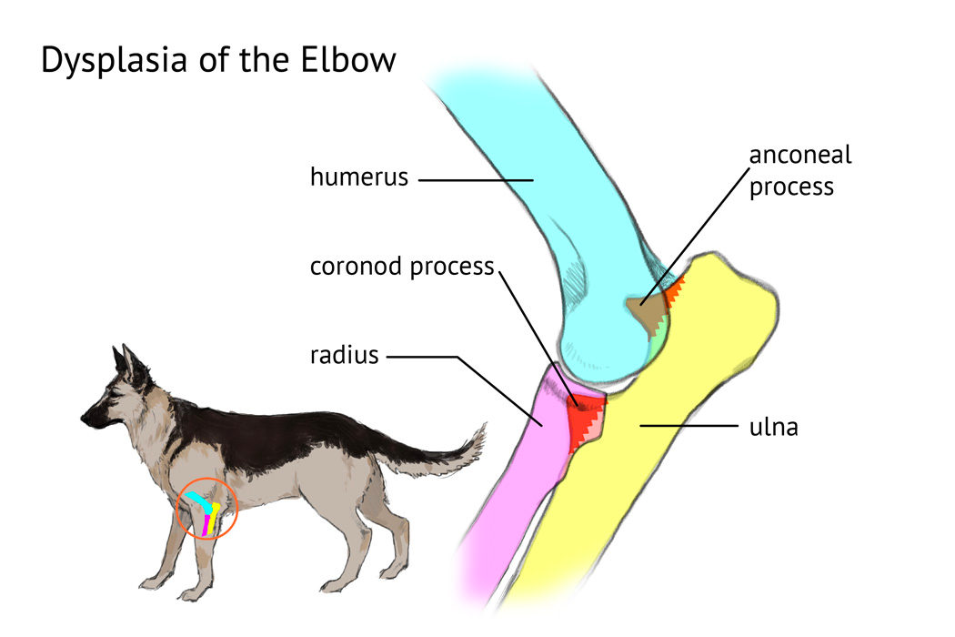
Elbow dysplasia, as the name suggests, relates to abnormal development of the elbow – especially the joints are affected. In major cases, both elbows are affected but unilateral elbow dysplasia cannot be ruled out. The ultimate result is severe arthritic change following elbow incongruities and bony chips or fragments.
The lameness in the leg arises from three different kinds of conditions, viz: Fragmentation of Medial Coronoid Process (EMCP) Osteochondritis Dissecans of Medial Humoral Condyle Non-joining of Anconeus Process (NJAP)
Within the first few months (fifth to twelfth) the puppies are vulnerable to this disease. In the body of a puppy, there are innumerable bone pieces with cartilage separating them. The formation of the long bone of the limbs, as the puppy grows, is interesting. The cartilage starts changing into the bone and all other minute bone pieces fuse to form an entire bone. For example, the ulna bone in the forearm is the combination of 4 bone pieces fused into one.
It will be interesting to delve further deep. The elbow joint is primarily composed of three bones – the radius, the humerus, and the ulna. For proper elbow functioning, all these three needs to fit in perfectly. The radius is the main weight bearer of the arm. The ulna acts as a lever arm for the extensor muscles of the elbow joint. In a normal elbow, the medial coronoid process of the ulna bone sits either at level or slightly below the surface of the radius. But in the case of the dysplastic elbow, the ulna position is above the level of the adjacent radius. It creates a step that is incongruous and thereby restricts the smooth transition between the two bones. In such a position the ulna is forced to take the weight-bearing force and logically the pressure increases on the medial coronoid process. The ultimate result is the fragmentation of the coronoid. Usually, the fragmentation replicates the size of the rice grain. Incomplete fragmentation results in the formation of fissures. Lesions might be preset on the humeral articular surface as well OCD (Osteochondritis Dissecans) or cartilage flap. Bone joints covered by cartilage fail to rub smoothly since the joint fluid starts leaking through the cracks, fractures, and fissures of the cartilage. This leakage causes pain because lubrication lessens.
Clinical Signs and Symptoms

The clinical symptoms of the disease are intermittent lameness and severe crippling, depending on the degree of the disease. The front legs of the dog show an abnormal posture and gait. The elbows are either stuck out or tucked in and the dog stands in a position of feet rotated outward. Swelling and grating are the other signs of the disease.
Puppies with elbow dysplasia are tagged as lazy because they prefer idling; even if they play it for a short duration. If both legs hurt the limp is occasional because the puppy manages its gait and stance by manipulation of elbows by shifting weight from one elbow to the other.
Breed of Dogs Affected by the Disease
German Shepherd dogs along with Bernese Mountain Dog, Golden Retrievers, Rottweilers, and Labrador Retrievers top the list of patients. Many other breeds of dogs are affected too – they are Saint Bernard, Mastiff, Springer Spaniel, Newfoundland, Chow Chow, Shar-Pei, Australian Shepherd, Terrier, and Shetland Sheepdog.
Is the Disease Genetic?
Just like hip dysplasia, elbow dysplasia depends on the genetic makeup of the body. Genetic pattern decides the development of cartilage degeneration (osteochondritis) and poor elbow joint. Hormones too play a part and it has been seen that male dogs are affected more than females. The reason is testosterone which enhances bone growth and ossification.
Hence an examination preceding the breeding procedure is helpful since the disease is genetically transmitted. Dogs with the best elbows can be chosen for the purpose of breed propagation. Genetic technology is best utilized to bring down the number of afflicted patients in the canine population.
How to Find the Disease?
Radiograph X-ray technique helps diagnose the incongruity and abnormalities of the elbow joint. It can detect the smallest fragments, lesions, and joint modifications. Magnetic Resonance Imaging or scanning is ideal for assessing lesions affecting the cartilage.
Treatment of Elbow Dysplasia
Elbow dysplasia is usually treated through the combined action of medicines and surgery. The objective is to relieve pain and maintain the proper functioning of the limb. Surgery is suggested before arthritic change starts taking place. There are two options for surgery with similar results– the traditional one and arthroscopy. Medical therapy includes weight management, a moderate amount of exercise, and pain-killing, anti-inflammatory medicines (NSAIDs). Natural vitamin supplements are gaining popularity because many pet owners do not like to take the risk of liver failure associated with NSAIDs. Since the disease is progressive, one can expect improvement but not normality. Managed hydrotherapy can prove to be an effective treatment for canine elbow dysplasia.
Treatment has to be coupled with environmental factors; diet being one of the crucial aspects. Overfeeding and excess calcium intake (hyper-calcification) are not desirable. A deficiency of Vitamins is one of the major causes of the dysplastic elbow in GSD and other canine breeds.

 Top 10 Best Sable German Shepherd Breeders in The United States